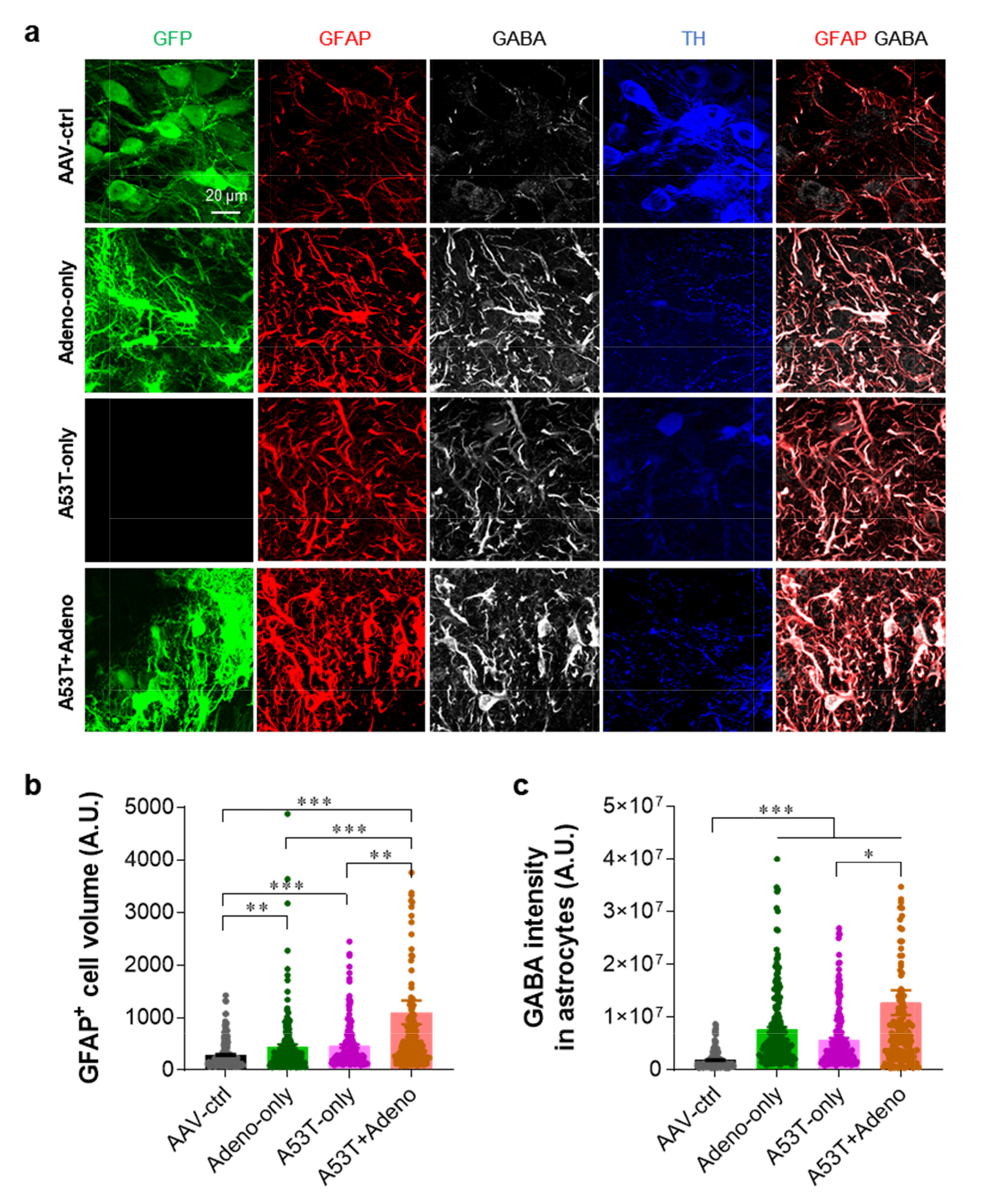
Fig. 3. Adenovirus injection exacerbates reactive astrogliosis. (a) Representative confocal images of SNpc tissues stained with TH, GFAP, and GABA. (b) Quantification of the volume of GFAP-positive (GFAP+) astrocytes in the SNpc. (c) Quantification of the integrated density of GABA in the GFAP-positive astrocytes. For all figures, mean±SEM; ns, non-significance, ***p<0.001 assessed by Kruskal-Wallis test with Dunn's multiple comparison test.
© Exp Neurobiol